
目次 at IgMin Research
We focus on enhancing knowledge exchange between disciplines to achieve rapid innovation.
Article Explorer
Evaluating Digital Imaging Technologies for Anogenital Injury Documentation in Sexual Assault Cases
The Role of Supplementation in Enhancing Recovery and Endurance among Fitness Trainers
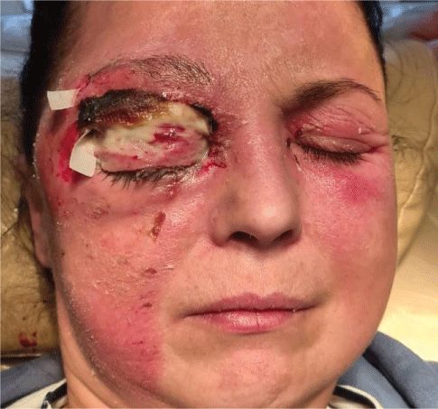
A Case of Facial Erysipelas with Necrosis of the Upper Eyelid
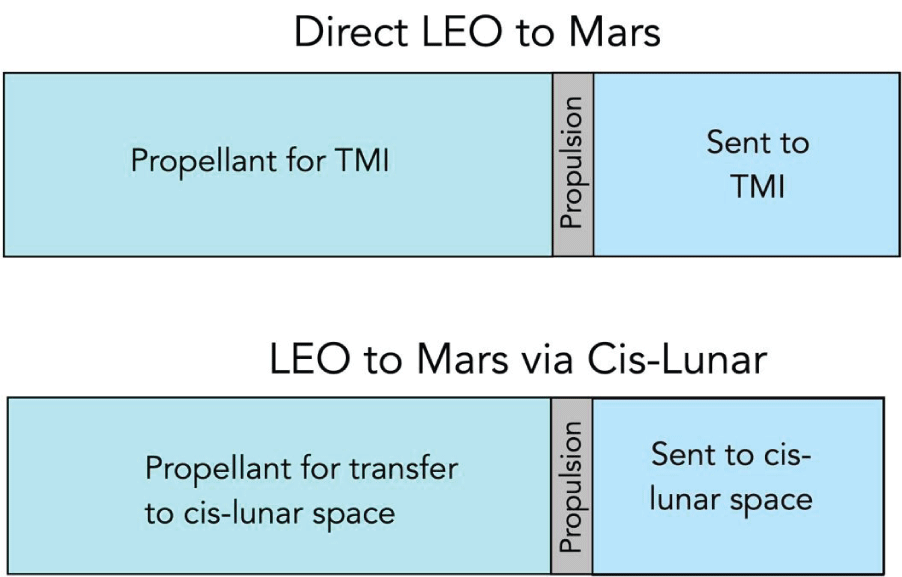
Lunar-Derived Propellants for Fueling Mars-Bound Spacecraft in Cis-Lunar Space
Risk Contagion Effects of Interconnected Manufacturing Enterprises
Exit Schreiner Collection
Use of Extraterrestrial Resources and Recycling Water: Curb Your Enthusiasm
Research Article
Evaluating Digital Imaging Technologies for Anogenital Injury Documentation in Sexual Assault Cases
Purpose: Photo-documentation is a critical component of care and an indispensable skill for forensic clinicians treating patients who have experienced violence and trauma. This retrospective study examines the frequency and nature of anogenital injuries identified through colposcope digital imaging in comparison to those detected using a high-resolution camera system.
Methods: This retrospective, before-and-after study evaluated genital injuries in all adult women (over 16 years old) who presented to a freestanding Nurse Examiner Clinic (NEC) following sexual assault during a 3-year period. The clinic is supervised by forensic clinicians trained in conducting medical-forensic examinations. In 2016-2017, all injuries were documented using the Cooper Surgical Leisegang© colposcope system, while in 2018, injuries were recorded exclusively with a high-resolution camera system. The primary outcome was the frequency of genital findings documented in sexual assault victims from each group.
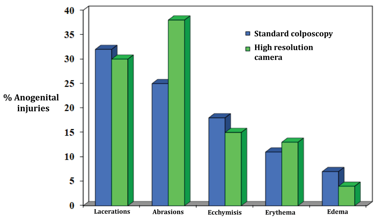
Results: A total of 367 women were evaluated in the "before" period and 180 in the "after" period. Both groups were similar in terms of demographics, assault history, time to examination, alcohol use, and the occurrence of genital injuries (76.1% vs. 74.9%, p = 0.76). However, patients examined with the high-resolution camera system had a significantly higher number of documented anogenital injuries (2.4 vs. 1.8, p < 0.001). This group also had more anogenital abrasions identified (51.1% vs. 27.0%, p < 0.001). The injury patterns between the two groups were not statistically different.
Conclusion: Accurate and reliable photo documentation are the key components of forensic medical documentation. Our findings indicate that the detection of anogenital injuries may differ based on the imaging system used.